Advertising disclosure: This page includes affiliate product links that may earn us a commission, as well as products from our own store.
A muscle can grow without every measurement describing that growth equally well. That matters when a headline turns a small change at one ultrasound site into a verdict on an entire exercise or training method. A 2026 comparison in 39 young men found that changes in quadriceps thickness did not reliably represent changes in MRI-based muscle size. The useful response is to ask exactly what was measured, rather than abandon research or book an expensive scan. Here is how to separate thickness, area and volume, read apparently conflicting findings, and build a progress record that keeps each number in its proper role.
What the 39-man comparison actually tested
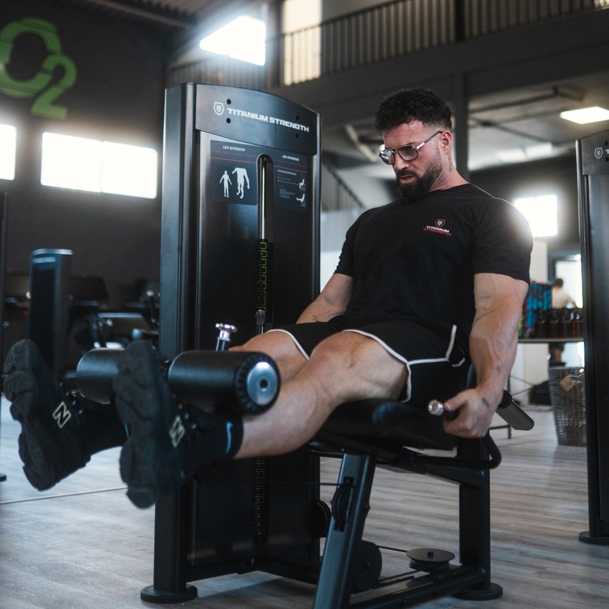
The men completed 15 weeks of lower-body resistance training, using knee extension, knee flexion and leg press three times weekly. Their dominant thighs were scanned before and after training. The comparison paired ultrasound muscle thickness with MRI measurements of maximum anatomical cross-sectional area and muscle volume, considering the quadriceps as a whole and its individual muscles.
For the overall quadriceps and two constituent muscles, vastus lateralis and vastus medialis, thickness changes underestimated MRI-based changes. No significant correlations linked thickness changes with either MRI outcome across the overall quadriceps or its constituent muscles. The result calls for caution when using thickness change as a stand-in for broader growth in this setting.
This was a measurement comparison, not a trial showing that one leg exercise builds more muscle than another. It does not supply a new winning program or a reason to replace your leg press. Keep the practical question aligned with the experiment: how well did one kind of measurement track another over a training period?
The same discipline applies when discussing a small exercise-selection study. Before treating an outcome as a complete muscle-growth score, check whether it represents a local measurement or a larger region. A study can be carefully conducted and still answer a narrower question than a social-media summary implies. That limitation belongs beside the result, not hidden after an absolute conclusion.
Thickness, area and volume are different questions
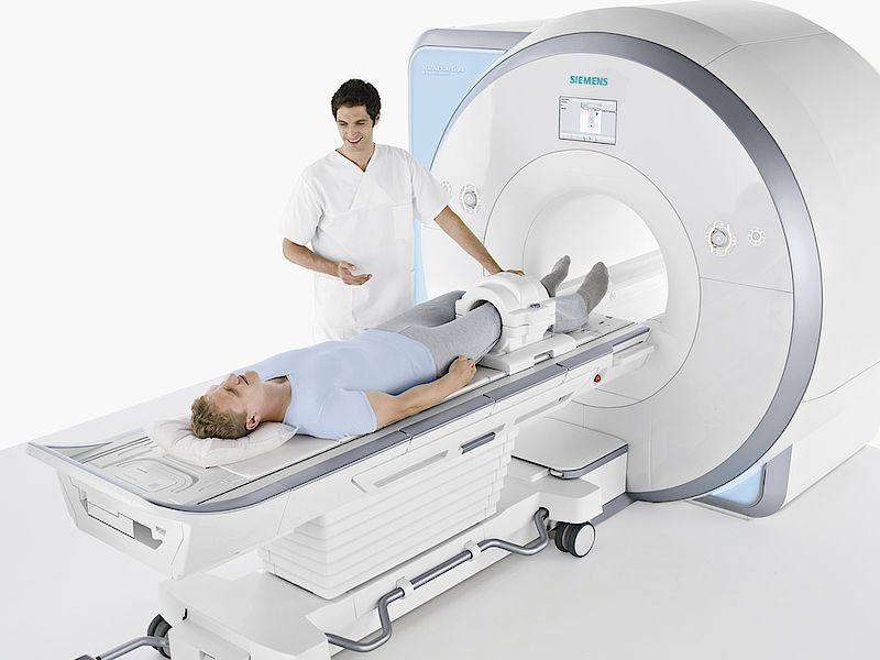
Thickness is a distance between boundaries at a measurement location. Cross-sectional area describes a two-dimensional slice. Volume describes three-dimensional size over an extent of the muscle. These words are not interchangeable, and a percentage attached to one should not quietly become a percentage attached to another.
Imagine comparing a single width measurement of a suitcase with the area of one face and its total storage volume. Each number describes something real, but one does not automatically tell you how the others changed. This is only a geometry analogy, not a model of muscle biology. Its value is in making the measurement question obvious before arguing about the result.
Even matching the instrument is insufficient. Two ultrasound studies may quantify different things: local thickness in one case and a reconstructed cross-sectional area in another. Likewise, a measurement at a selected point is different from searching for the largest slice. Read the outcome label and location before placing results side by side.
For example, a claim that an exercise increased local thickness by a given percentage should remain a claim about that measurement. You need additional evidence before describing an equal percentage increase in the entire muscle's volume. A smaller number from another method is not automatically worse training, and a larger number is not automatically a superior exercise. The methods must be comparable before the ranking becomes meaningful.
Why this does not make all ultrasound research worthless
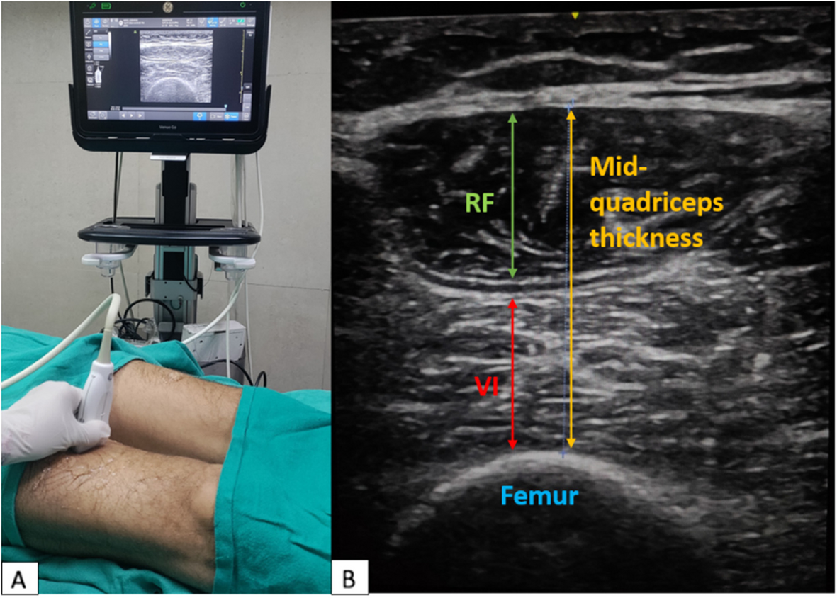
An earlier investigation in 14 young men compared ultrasound-derived and MRI-derived changes in mid-thigh vastus lateralis cross-sectional area. After ten weeks of unilateral training, the two approaches showed similar average increases and correlated changes. That finding concerned area measured with a particular protocol. It did not establish that every thickness measurement predicts whole-muscle volume.
The sensible reading is conditional: an instrument can be useful for a defined outcome under a defined procedure. A favorable validation of one technique does not validate every application of the same technology. A disappointing comparison of another technique does not erase every useful application either.
Reliability and validity also deserve separate questions. Can a procedure produce repeatable numbers? And does the number represent the outcome you actually want to know? Repeatedly measuring the same location carefully is valuable, but consistency alone cannot turn a local distance into total volume. Conversely, a theoretically relevant outcome is less helpful when the procedure is too variable to track.
When research findings seem to disagree, begin with the methods before inventing a dramatic reversal. Compare the measured quantity, muscle, site, participants and training period. Do not demand that every paper reach an identical conclusion from a different question. Keep uncertainty attached to the particular comparison instead of declaring an entire body of training research either proven or debunked.
Repeatability starts with the setup
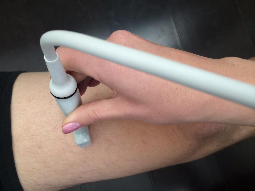
Ultrasound acquisition involves more than putting a probe somewhere on the thigh. Measurement procedures specify anatomical landmarks, probe orientation and image capture. Research examining technique has also shown why pressure matters: compressing tissue can alter the thickness being recorded. Those details explain why a casual repeat measurement may be harder to interpret than its precise-looking number suggests.
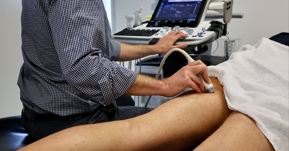
A professional scanning protocol may use a tape to locate a position, then adjust the probe relative to visible muscle boundaries. That tape is locating a scan site; it is not measuring muscle volume. Keep each step's purpose clear. A picture of a probe does not tell you which imaging outcome was ultimately calculated.
The practical parallel for ordinary lifting is to standardize the comparison you can control. If you record thigh circumference, use the same location, posture and tape technique each time. Avoid comparing a tightly pulled tape after a workout with a relaxed measurement on another day. Write down changed conditions instead of pretending every reading is equally comparable.
A tape measure remains a circumference measurement around the limb, including more than muscle. It is not a substitute for a validated imaging method. That does not make it useless for a personal record; it means you should describe it honestly. If the number changes, consider it alongside other observations rather than announcing an exact amount of new muscle tissue.
Apply the same thinking to progress photographs. Similar camera position, lighting, stance and distance make a comparison easier to judge. They still do not transform a photograph into a laboratory scan. Standardization improves your personal record while leaving the limits of the method intact.

Equipment Worth Exploring
Explore equipment, accessories and gear from our carefully selected range.

Leg Extension/Curl BCO-780
Leg extension and lying leg curl combo with plate loading, band pegs and a cable-loading option.

Seated Leg Combo BCO-680
Adjustable seated leg extension and leg curl machine with band pegs for added resistance.

Leg Press/Hack Squat BPR-790
Convertible 45-degree leg press and hack squat machine with a broad footplate and band pegs.

Compact Leg Press CLP-700
Compact reverse-style leg press with a moving seat carriage and an adjustable footplate.


GymPin Squat Wedges
Non-slip 15-degree squat wedges help elevate the heels for deeper quad-focused squats.

Studio V-Squat
Plate-loaded V-squat machine for supported lower-body strength training.

Rubber Hex Dumbbells
Rubber-coated hex dumbbell pairs with a stable shape for home and commercial strength training.

Use a dashboard instead of one magic growth number
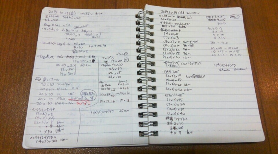
Start by deciding what your training record needs to answer. If the question is whether you are getting stronger at a leg extension, record that exercise's performance. If the question concerns appearance, use consistently taken photographs. If you track circumference, keep it in its own column. Combining observations is different from pretending that every observation measures the same thing.
For a simple four-week record, note the machine, seat and pad settings, load, repetitions and range of motion for your main lower-body exercises. Review repeated sessions rather than one unusually good set. A heavier leg press performed with a shorter movement is not a clean comparison with the previous version, even when the weight number looks impressive.
Keep a separate place for bodyweight trends, photographs and any circumference readings you choose to collect. These can provide context for interpreting your experience. They cannot establish an exact MRI-equivalent change in quadriceps volume. Avoid turning the dashboard into a homemade equation that converts strength, scale weight or tape readings directly into kilograms of muscle.
Regular training still matters more than endlessly replacing measurement tools. Build a program you can sustain, track it consistently and adjust it for your goals and recovery. Changing exercises, volume and measurement procedures simultaneously makes it difficult to understand what happened. When you make a deliberate change, document it and establish a new comparison.
Finally, apply the same restraint when reading the next growth headline. Identify the measured outcome, look for a meaningful comparison and keep conclusions within that scope. The lesson is not that progress cannot be measured. It is that a useful number needs a clear definition, a repeatable procedure and an interpretation that does not promise more than the measurement can deliver.

Funny Gym Shirts & Workout Gear
Funny gym shirts, tanks, hoodies and workout gear for lifters who like training hard.

Quadzilla Massive Quads Sweater Leg Day Squatting T-Shirt
Keep things casual with a little “Quadzilla” attitude.

Lift Smart Brain Weight Plate Mind Muscle Connection 45 T-Shirt
For laid-back days and lifting humor: “Lift Smart Brain Weight Plate”.

Pencilneck Science Based Lifting Gym | Funny Workout De Sticker
Stick a little “Pencilneck Science Based Lifting” humor on your favorite gear.

Bro Science Swole Scientist | Bodybuilding & Powerlifti Sticker
Stick a little “Bro Science Swole Scientist” humor on your favorite gear.


Musclenomics Muscle Graph : The Ultimate Meme Shirt & T-Shirt
Make gym days more fun with “Musclenomics Muscle Graph” style.

Progress Stoic Bodybuilder Greek God Marble Sculpture T-Shirt
Heavy lifts, good laughs, and a little “Progress Stoic Bodybuilder Greek God Marble” energy.

Bro Science Element Chemical T-Shirt
Show some bodybuilding attitude with a playful “Bro Science Element” tee.

Never Skip Leg Day Chicken Legs Meme & Bros Tank Top
Bring “Never Skip Leg Day” humor to your next lifting session.

Gym Nerd Pixel Lifting Meme Workout Bodybuilding Powerl Hoodie
Make your off-duty look more fun with “Gym Nerd”.