Can lifting weights change an aging brain? A brain scan cannot tell us whether someone remembers where they left their keys, but it can reveal differences in the microscopic environment of pathways connecting brain regions. That distinction matters when exercise research attracts headlines. A 2026 randomized trial suggests that strength and balance practice can influence certain white-matter measurements in older adults. The result is encouraging, but it is not evidence that dumbbell workouts reverse dementia or make a brain biologically younger.
What the three-month trial actually tested
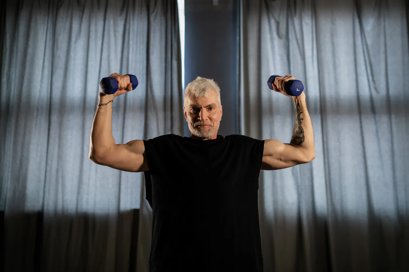
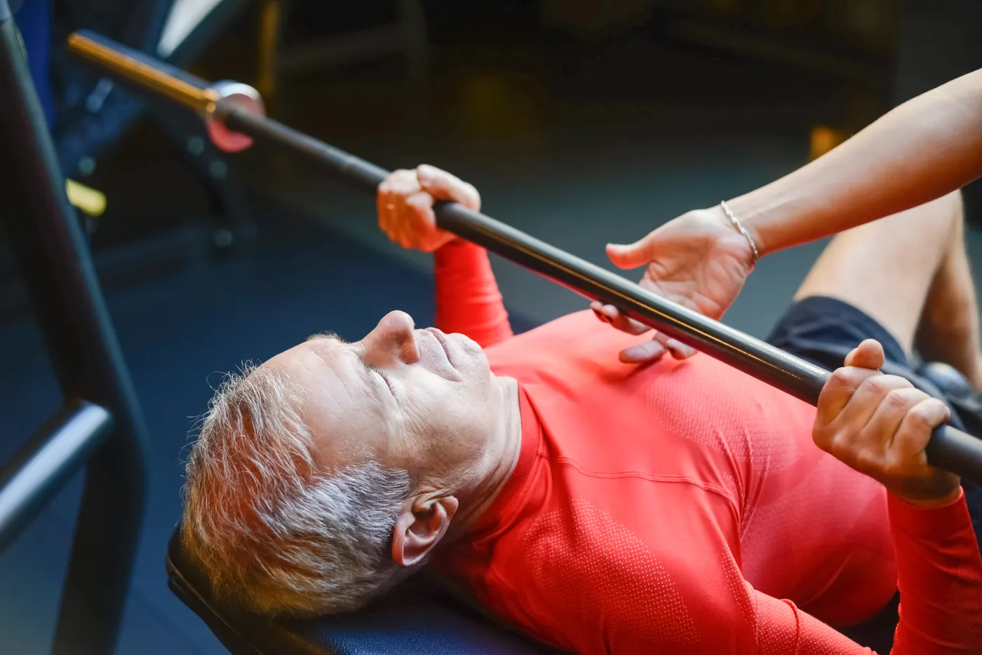
Researchers studied 60 adults aged 64 to 82, with an average age of about 71. Participants were assigned to strength training, balance training or a passive control group. The active groups trained for three months. Investigators used several MRI-based methods to examine changes in brain tissue, especially white matter, the bundles of nerve fibers carrying signals between regions.
This design is more informative than comparing lifelong exercisers with people who rarely move. Random assignment reduces some pre-existing differences between groups and helps researchers ask whether measurable changes emerge over a defined period. The control group provides context for changes that might happen with aging or repeated testing even without exercise.
However, the outcomes must be read carefully. The main findings concern imaging markers, not a memory test, a dementia diagnosis or a measured improvement in daily thinking. A scan-based difference may suggest that the nervous system responds to training, but its practical importance requires separate evidence. The useful interpretation is that physical training might affect the aging brain’s tissue environment over a relatively short period. That is a worthwhile scientific finding without turning it into a miracle claim.
What changed in the brain’s white matter?
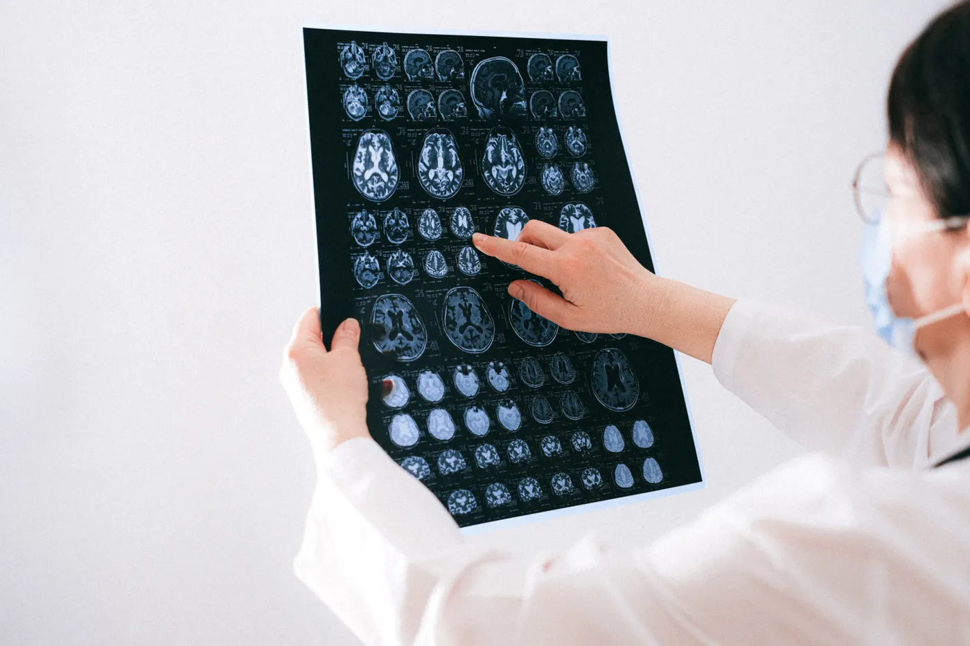
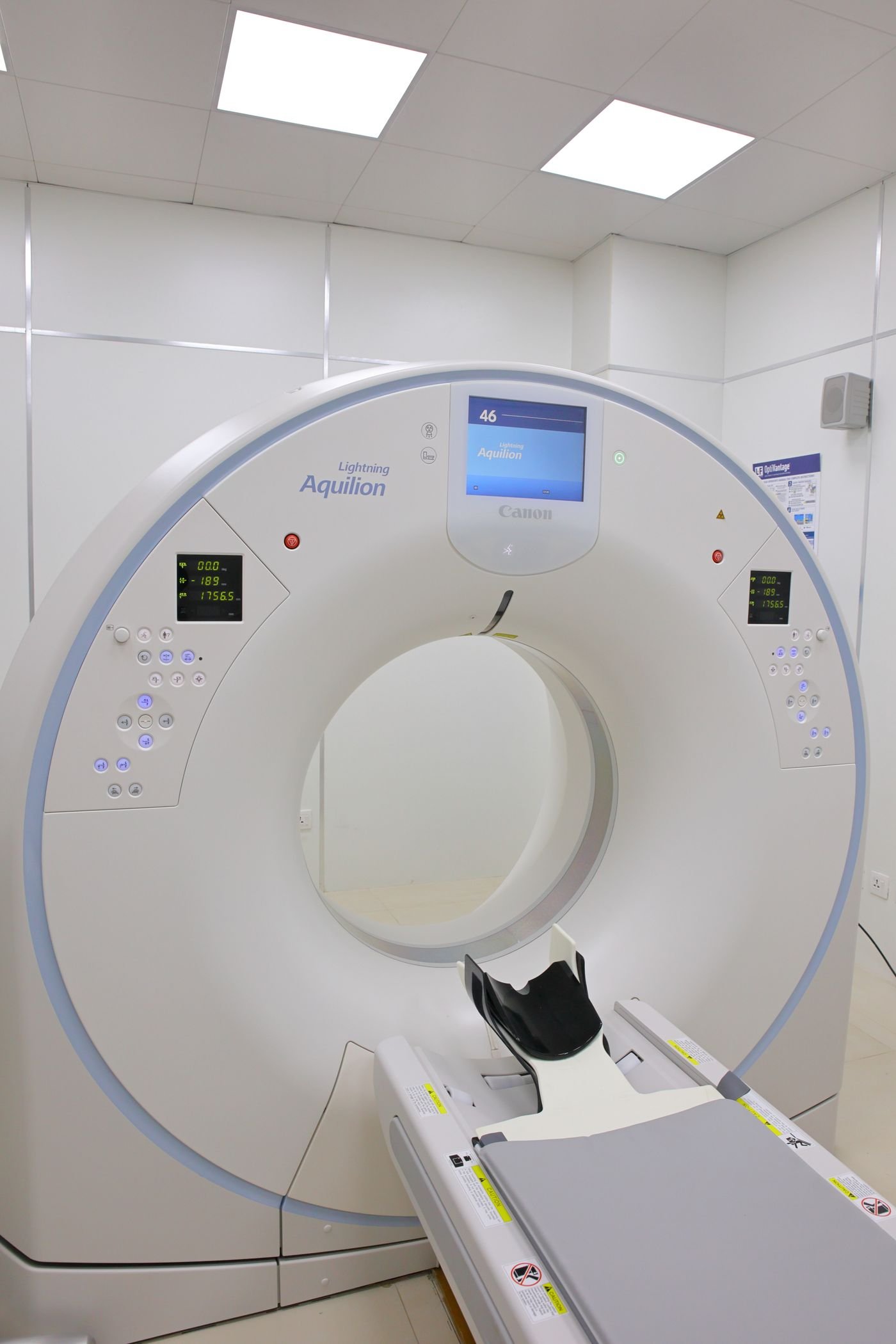
White matter is often described as the brain’s wiring. It contains long connections between regions, supported by myelin and other tissue structures. The study used diffusion MRI: rather than watching electrical impulses directly, the scanner estimates how water molecules move through brain tissue. Statistical models then translate those patterns into measurements that may reflect tissue organization.
In the strength-training group, researchers reported changes in extra-axonal axial diffusivity in bilateral inferior fronto-occipital and longitudinal fasciculi. Those technical names describe long pathways connecting different areas of the brain. The pattern was interpreted as consistent with microstructural remodeling, not as proof that new nerve fibers grew or that participants became cognitively faster.
Both active groups also appeared to mitigate a decline in axonal water fraction seen in controls, including parts of the corpus callosum and right posterior corona radiata. Axonal water fraction is a model-derived MRI measurement. It is not a direct count of surviving neurons, and it cannot independently prove that myelin was rebuilt. A difference in one measurement should not be converted into a sweeping claim about the entire brain.
The word change can describe a meaningful adaptation or a measurement whose long-term consequences remain uncertain. This trial supports the possibility of physiological adaptation but cannot settle its clinical importance. It gives scientists a reason to study strength and balance training further; it does not provide a new diagnostic test for brain aging. That nuance matters when translating an interesting laboratory result into everyday advice.
Equipment Worth Exploring
Explore equipment, accessories and gear from our carefully selected range.


FID Bench MAX
Adjustable bench offering flat, incline, and decline positions + adjustable headrest.
VOLTRA
Compact digital cable trainer with smooth, adjustable resistance.

Go-Tone Pro
Portable digital cable trainer with up to 66 lb (30 kg) of adjustable resistance.


Strength and balance training did not produce identical signals
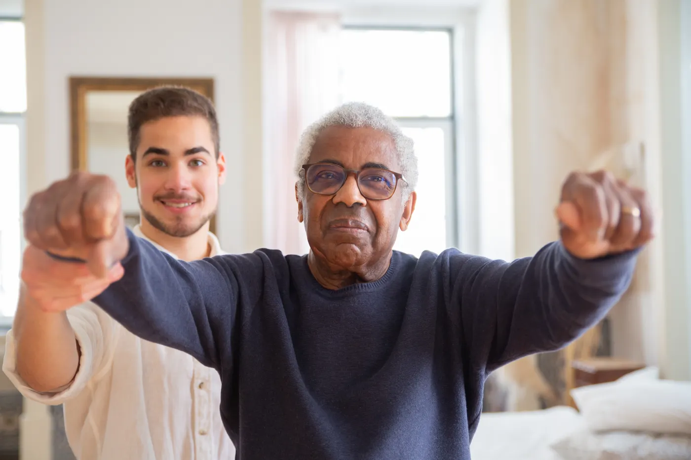
One useful feature of the experiment is that it compared two genuinely different approaches. Strength work challenges muscles to produce force against resistance. Balance practice challenges the nervous system to organize posture and movement while the body stays stable or changes position. Both demand coordination, but they do not stress the body in identical ways.
In the MRI results, both training groups showed preservation of certain white-matter measures compared with the passive control group. The strength group additionally showed changes in diffusion measures within long association pathways. That does not automatically mean strength training was better for brain health. Different metrics answer different questions, and the reported results do not establish a universal ranking between the programs.
For someone building a practical routine, this argues against treating resistance and balance exercise as competitors. Strength can make it easier to rise from a chair, carry groceries or control a step. Balance practice can improve confidence and skill in standing, turning and reaching. Those functional reasons already justify doing both, even if later research finds that the imaging effects are smaller or less durable than hoped.
A separate 2026 systematic review of balance interventions in older adults reported a small average improvement in pooled cognitive outcomes. That is useful context, but it should not be combined with this MRI trial as though they measured the same thing. One examined performance on cognitive tests; the other primarily examined tissue-related scan metrics. The distinction prevents us from overstating what any one result means.

Funny Gym Shirts & Workout Gear
Funny gym shirts, tanks, hoodies and workout gear for lifters who like training hard.

Mind Muscle Perfection Brain and Biceps Gym for Science T-Shirt
Show some gym-day personality with “Mind Muscle Perfection Brain and Biceps”.

Lift Smart Brain Weight Plate Mind Muscle Connection 45 Tank Top
Add some lifting attitude to training with “Lift Smart Brain Weight Plate”.

Strong Inside And Out Shirt Weights And Brains Meme T-Shirt
Rep “Strong Inside And Out” humor in and out of the gym.

Gym Is My Happy Place Happy Weights Emoji Weight Plate Hoodie
Show off your powerlifting humor in a laid-back “Gym Is My Happy Place” hoodie.

Science Based Lifting Rick And Morty Decal Water Sticker
Stick a little “Science Based Lifting” humor on your favorite gear.

Physical Mass Einstein Tee
Show some bodybuilding attitude with a playful “Physical Mass” tee.

Beatles Lift Together – Lift Together Hooded Pullover Hoodie
Keep it laid-back with a little “Beatles Lift Together” attitude.

Gymhead Lifting Is On My Mind Decal Skull Meme Sticker
Bring “Gymhead” attitude to your water bottle or laptop.

Progress Stoic Sigma Greek God Marble Sculpture – T-Shirt
Rep “Progress Stoic Sigma Greek God Marble” humor in and out of the gym.

Lifting Is My Therapy Gymbros, & Meme Shirt & Gains T-Shirt
Rep “Lifting Is My Therapy” humor in and out of the gym.
What this research does not prove
The trial was randomized, which is a strength, but 60 participants is still a modest sample. Three months may reveal adaptations and is far too short to establish whether they persist for years. The participants were aged 64 to 82; the findings cannot automatically be extended to younger lifters, people with advanced neurological disease or everyone with different medical conditions.
Most importantly, the abstract does not report that participants improved their memory, decision-making or everyday independence because of the MRI changes. It does not demonstrate prevention of Alzheimer’s disease, reversal of dementia or a reduction in a validated biological brain-age score. Diffusion metrics are not interchangeable with any of those outcomes.
Other studies provide context. A separate randomized resistance-training experiment published in 2026 used functional connectivity and brain-age prediction models and reported changes in those particular outcomes. Another small trial involving water-based resistance training in older women reported structural and blood-marker differences after 12 weeks. These are not identical replications: populations, exercise protocols, imaging methods and endpoints differ substantially.
When several studies point in a similar direction, confidence increases that movement and brain biology are connected. It does not eliminate uncertainty about the best dose, ideal exercises or clinical importance of an individual scan measurement. Nor does it show that lifting is a substitute for medical treatment, adequate sleep, social connection or other aspects of healthy aging. Anyone promising guaranteed brain rejuvenation from one machine or exercise is going well beyond the evidence.
Why lifting might influence neural tissue
Resistance exercise is not just a local event inside the biceps or quadriceps. Every repetition requires the nervous system to recruit muscle fibers, coordinate joints, regulate breathing and adjust force as fatigue builds. Over time, training can change how efficiently a person performs a movement. It also affects circulation, metabolic demand and the body’s broader physiological environment.
These processes make it biologically plausible that training influences the brain. But plausibility is not a proven mechanism. The MRI study did not isolate whether its tissue-related signals came from vascular regulation, myelin properties, neural activity, inflammation, learning a movement or several factors acting together.
Consider an analogy: a stronger engine can make a vehicle more capable, but a photograph of the engine cannot tell you whether the driver has become more skilled. Likewise, a white-matter signal may be encouraging without demonstrating that concentration or memory improved. Good science keeps those questions separate instead of allowing an exciting image to answer everything.
The practical implication is not to chase an MRI number. Choose sustainable training that improves outcomes people can feel and measure: safer movement, stronger legs, better grip, easier stair climbing and confidence in daily tasks. Brain-related effects may be an additional benefit, but functional gains are already valuable. There is no need to make the routine complicated simply because the neuroscience sounds complicated.
Equipment Worth Exploring
Explore equipment, accessories and gear from our carefully selected range.


GymPin Squat Wedges
Non-slip 15-degree squat wedges help elevate the heels for deeper quad-focused squats.

Safety Squat Handles
Barbell squat handles that reduce shoulder positioning demands during high-bar squats.
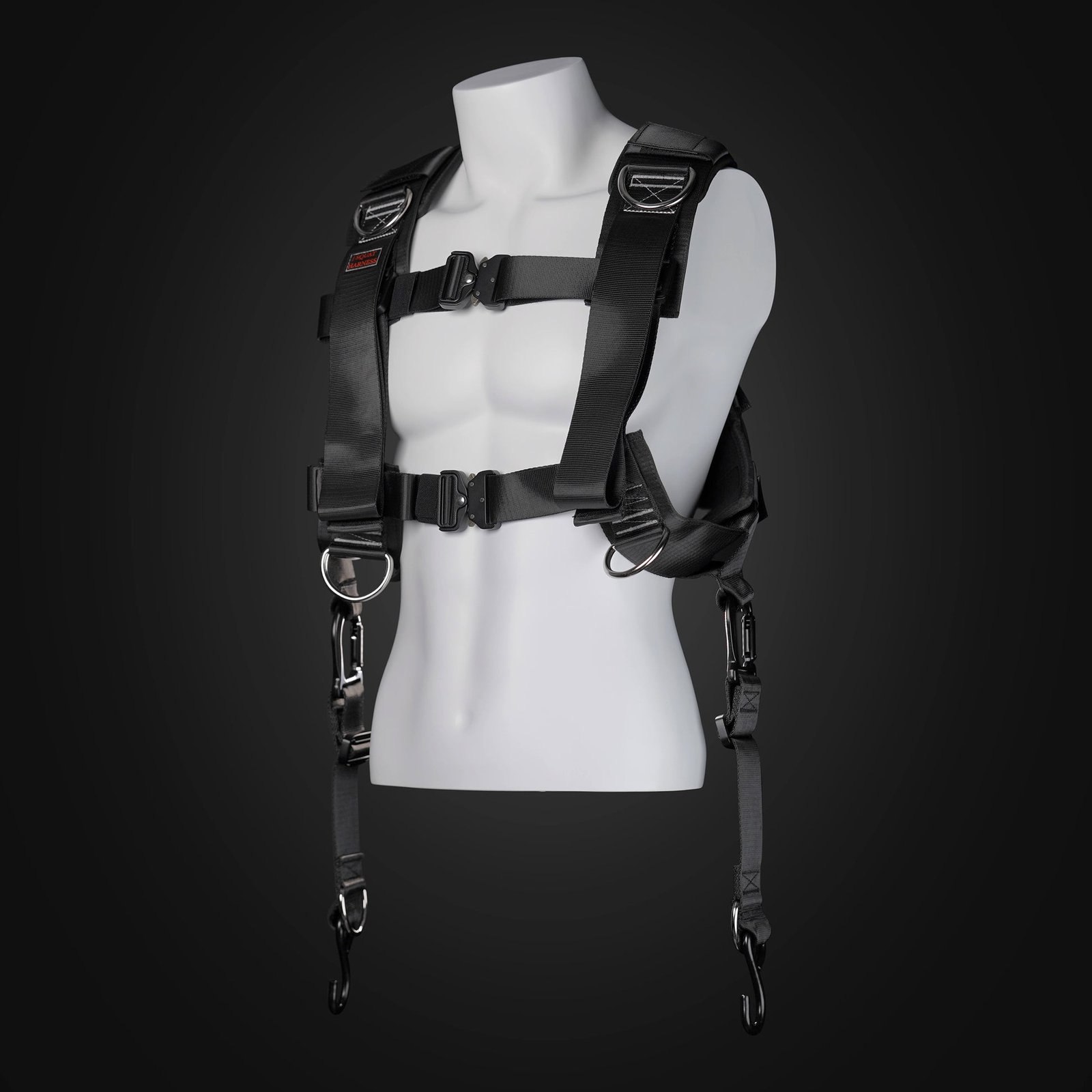
Squat Harness
Wearable harness for hands-free cable squats, sled work, and lower-body training.


HUSLA Leg Extension Pad
Pad attachment designed to increase usable range of motion on compatible leg-extension machines.


A realistic two-day strength routine for older adults
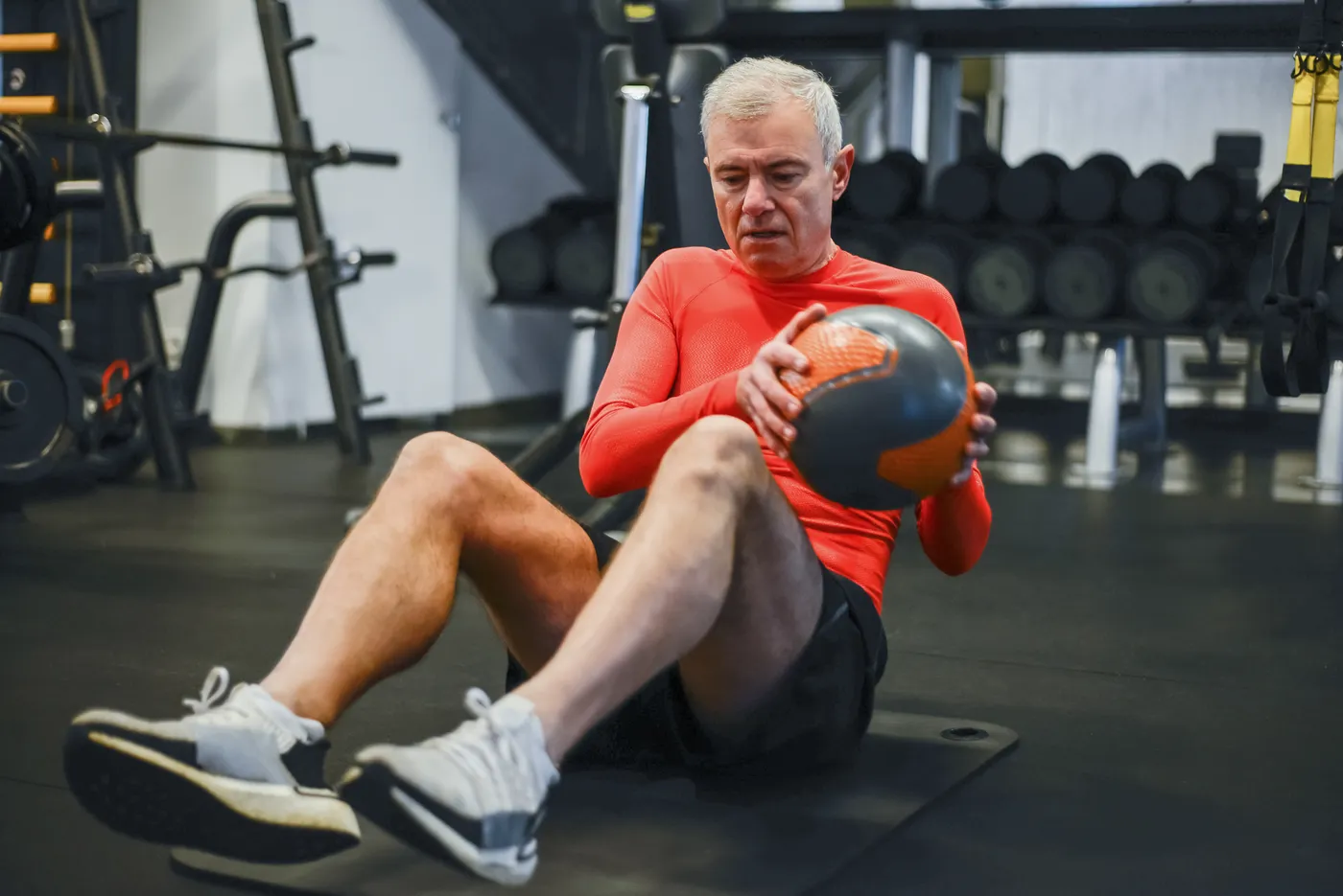
A sensible starting point is two nonconsecutive full-body sessions per week, allowing roughly 30 to 45 minutes for each. This is a general practical example, not the exact protocol used in the MRI trial. Begin with comfortable walking and easy joint movements. Select resistance that permits smooth repetitions without breath-holding or loss of balance.
Movement one: sit-to-stand or supported squat. Do two sets of six to ten repetitions using a stable chair as a reference. Adjust chair height or hold a light weight only when the movement is secure. This practices standing up without turning every workout into a maximal squat test.
Movement two: hip hinge. Try two sets of eight to twelve repetitions with a light dumbbell or an unloaded movement pattern. Keep the weight close and move through a comfortable range. A controlled hinge supports picking objects up while maintaining awareness of the hips and trunk.
Movement three: row. Use a band or cable for two sets of eight to twelve. A supported row may be preferable if standing balance is limited. Movement four: push. Choose a wall push-up, elevated push-up or seated dumbbell press for two sets of six to twelve. Movement five: carry. Hold light weights and walk for 20 to 40 seconds if gait is steady, or practice a supported standing hold.
Rest as needed, particularly after movements that challenge breathing or balance. When all repetitions feel controlled for several sessions, add one or two repetitions or a small resistance increase. The goal is gradual improvement, not exhaustion. People with major balance difficulties, recent falls or unstable medical conditions should adapt the plan with an appropriate clinician or therapist rather than copying a generic routine. No single piece of equipment is essential to the basic movement patterns.
Functional Fitness Shirts & Workout Gear
Functional-fitness shirts, tanks and workout gear for athletes who train strength, conditioning and everything in between.

Vive La Resistance Bands – Resistance Bands Warmup Mobi T-Shirt
Rep “Vive La Resistance Bands” humor in and out of the gym.

Atlas Kettlebell Earth Meme Greek God Top T-Shirt
Make gym days more fun with “Atlas Kettlebell Earth Lifter” style.

Lift Together Grow Stronger for Gym Buddies and Lifting Tank Top
Bring “Lift Together Grow Stronger” humor to your next lifting session.

Break The Stereotype | Feminist Workout Sweater For Str T-Shirt
For laid-back days and gym-girl humor: “Break The Stereotype”.

Gym Is My Happy Place Happy Kettlebell Emoji Hoodie
Show off your gym-girl humor in a laid-back “Gym Is My Happy Place” hoodie.

Kettlebell Heartbeat Muscle Shirt Athletic Vest Workout Tank Top
Add some lifting attitude to training with “Kettlebell Heartbeat”.

Live Love Lift Hooded Pullover Workout Gymbro Apparel Hoodie
Keep it laid-back with a little “Live Love Lift” attitude.

Kettlebell Heart X-Ray – Valentine's Workout Vest Muscl Tank Top
Train hard, laugh a little, and rep “Kettlebell Heart X-Ray”.

Super Kettlebell Crewneck Pullover Sweater Kettlebell T T-Shirt
Add a playful “Super Kettlebell” touch to your casual rotation.

Hybrid Athlete – Strength & Endurance Gym Decal for Run Sticker
Put “Hybrid Athlete” gym humor on your laptop, bottle, or gym gear.
Add balance practice—and track outcomes that matter
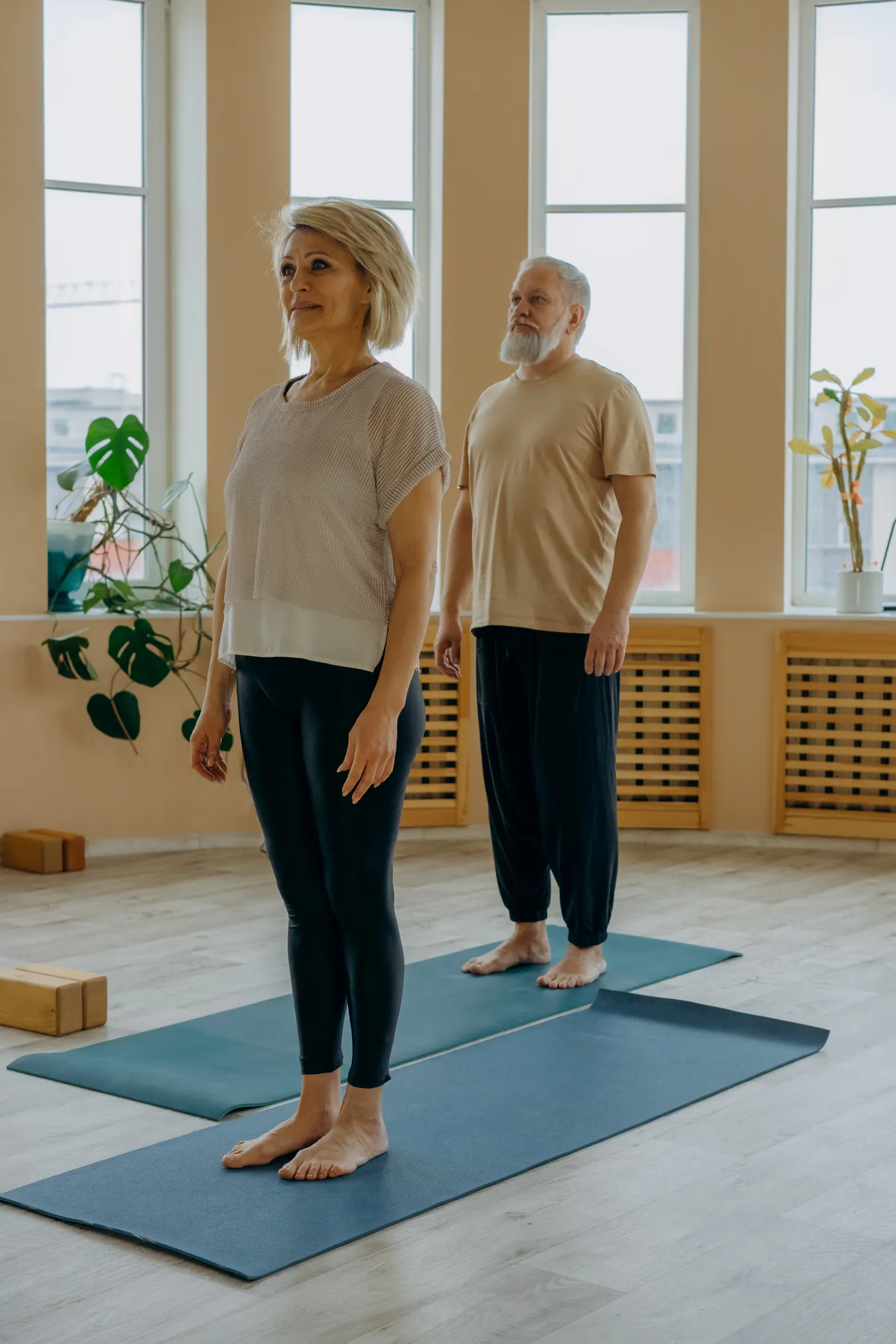
Balance can be practiced in short blocks between strength sessions. Three sessions of five to ten minutes may be easier to sustain than one elaborate workout. Start beside a sturdy counter or rail where support is immediately available. Practice a narrow stance, controlled weight shifts and slow side steps. If safe, progress toward a tandem stance or brief single-leg holds without sacrificing the ability to catch yourself.
Make tasks slightly harder only after they become repeatable. Closing the eyes, standing on unstable surfaces or turning quickly can increase fall risk, so those are not default progressions for unsupervised beginners. A clear floor, supportive shoes and reachable hand support matter more than sophisticated balance equipment. A steady routine is better than a dramatic challenge that someone is afraid to repeat.
Keep a simple log for six to twelve weeks. Note controlled chair rises, resistance used for rows, confidence carrying groceries and whether a supported balance hold becomes steadier. Record missed sessions without treating them as failure; consistency over months matters more than a perfect week. Include walking or another comfortable aerobic activity alongside strength work where practical.
These measures cannot reveal whether your corpus callosum changed on an MRI. They can show whether training improves everyday capacity, which is a compelling reason to continue. The 2026 imaging trial adds an intriguing possibility: strength and balance exercise may influence the aging nervous system in ways not immediately visible in the mirror. It does not replace the simpler conclusion that moving regularly, progressively and safely is worthwhile.